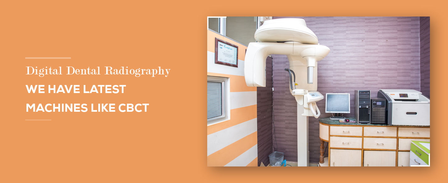
Digital Dental Radiography
What is Dental Radiograph?
Radiographs are commonly called X-rays. Dentists use radiographs for many reasons: to find hidden dental structures, malignant or benign masses, bone loss, and cavities. A radiographic image is formed by a controlled burst of X-ray radiation which penetrates oral structures at different levels, depending on varying anatomical densities, before striking the film or sensor. Teeth appear lighter because less radiation penetrates them to reach the film. Dental caries, infections and other changes in the bone density, and the periodontal ligament, appear darker because X-rays readily penetrate these less dense structures. Dental restorations may appear lighter or darker, depending on the density of the material.
What are Digital Dental Radiographs?
Digital radiography is a type of X-ray imaging that uses digital X-ray sensors to replace traditional photographic X-ray film, producing enhanced computer images of teeth, gums, and other oral structures and conditions.
What are various types of Digital Dental Radiographs?
Digital dental radiographs can be taken from inside (intraoral) or outside (extraoral) the mouth. Intraoral X-rays, the most commonly taken dental X-ray, provide great detail and are used to detect cavities, check the status of developing teeth and monitor teeth and bone health. Extraoral X-rays do not provide the detail of intraoral X-rays and are not used to identify individual tooth problems. However, they are used to detect impacted teeth, monitor jaw growth and development, and identify potential problems between teeth, jaws and temporomandibular joints (TMJ), or other facial bones.
Types of intraoral X-rays include:
- Bitewing X-rays which are taken with the patient biting down on film, show details of the upper and lower teeth in one area of the mouth. Each bitewing shows a tooth from its crown (top) to about the level of the supporting bone. Bitewing X-rays are used to detect decay between teeth and changes in bone density caused by gum disease, as well as to determine the fit of dental crowns or restorations, and the marginal integrity of tooth fillings.
- Periapical (limited) X-rays show the whole tooth from the crown to beyond the root tips to the supporting bone in one area of either the upper or lower jaw. Periapical X-rays are used to detect root structure and surrounding bone structure abnormalities. Showing bone loss around each tooth, periapical X-rays aid in treating conditions such as periodontitis, advanced gum disease, and detecting endodontic lesions (abscess).
Types of extraoral X-rays include:
- Panoramic (OPG) X-rays which require a machine that rotates around the head, show the entire mouth, including all the teeth in the upper and lower arch, in one image. Panoramic X-rays are used to plan treatment for dental implants, detect impacted wisdom teeth and jaw problems, and diagnose bony tumors and cysts. Panoramic films are used for forensic and legal purposes to identify otherwise unrecognizable bodies after fires, crashes or other fatalities.
- Multi-slice computed tomography (MCT) shows a particular layer or “slice” of the mouth while blurring all other layers. This type of X-ray is useful for examining structures that are difficult to see clearly.
- Cephalometric projections which show the entire head, help examine teeth in relation to a patient’s jaw and profile. Orthodontists, specialists in aligning and straightening teeth, use cephalometric projections to develop their treatment plans.
- Cone beam computerized tomography (CBCT) shows the body’s interior structures as a three-dimensional image. CBCT — often performed in a hospital or imaging center, but increasingly being used in the dental office — is used to identify facial bone problems, such as tumors or fractures. CT scans also are used to evaluate bone for dental implant placement and difficult tooth extractions to avoid possible complications during and after surgical procedures.
The CBCT, which requires up to eight times more radiation than panoramic radiographs, does not slice images. Instead, its cone-shaped beam scans both the upper and lower mouth areas at once. The data is captured by a two-dimensional array and creates high-resolution images, which are then combined to form a 3-D image of the bony structures.
What are benefits of Digital Dental Radiography?
Benefits of digital dental radiographs compared to traditional dental X-rays include the following:
- Digital radiographs reveal small hidden areas of decay between teeth or below existing restorations (fillings), bone infections, gum (periodontal) disease, abscesses or cysts, developmental abnormalities and tumors that cannot be detected with only a visual dental examination.
- Digital radiographs can be viewed instantly on any computer screen, manipulated to enhance contrast and detail, and transmitted electronically to specialists without quality loss.
- Early detection and treatment of dental problems can save time, money and discomfort.
- Digital micro-storage technology allows greater data storage capacity on small, space-saving drives.
- Dental digital radiographs eliminate chemical processing and disposal of hazardous wastes and lead foil, thereby presenting a “greener” and eco-friendly alternative.
- Digital radiographs can be transferred easily to other dentists with compatible computer technology, or photo printed for dentists without compatible technology.
- Digital sensors and PSP (photostimulable phosphor) plates are more sensitive to X-radiation and require 50 to 80 percent less radiation than film. This technology adheres to the ALARA (As Low As Reasonably Achievable) principle, which promotes radiation safety.
- Digital radiograph features, including contrasting, colorizing, 3-D, sharpness, flip, zoom, etc., assist in detection and interpretation, which in turn assist in diagnosis and patient education. Digital images of problem areas can be transferred and enhanced on a computer screen next to the patient’s chair.
- Digital dental images can be stored easily in electronic patient records and, sent quickly electronically to insurance companies, referring dentists or consultants, often eliminating or reducing treatment disruption and leading to faster dental insurance reimbursements.
We at Gulab dental hospital are fully equipped with digital dental radiography units which includes:
- Kodak 5100 RVG sensor from Kodak Dental imaging system (KDIS), France
- X-MIND DC x-ray unit by Satelac
- Cranex Novus-e panoramic imaging unit from Soredex
- CS 9000 3D Cone beam computed tomography machine (CBCT) from Carestream Dental
- Dryview 5700 Laser Imager
